3D Data Workflows and Tutorials
|
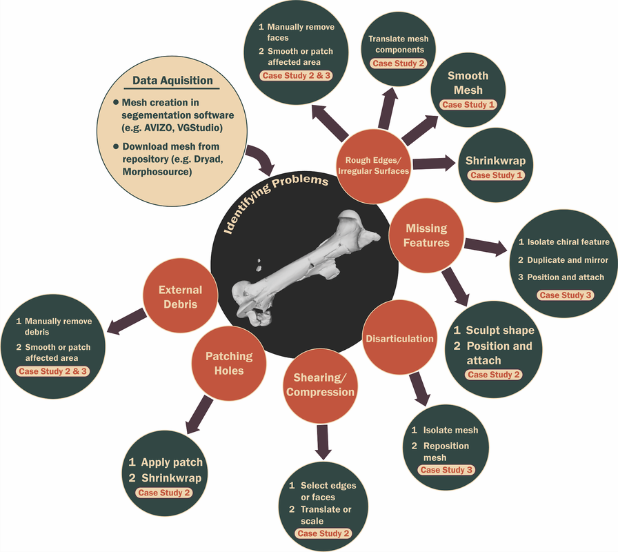
No gatekeeping here! The workflow used to make most of the 3D reconstructions featured on this website was recently published in PLoS ONE. An accompanying series of step-by-step tutorials takes users of any experience level through the basics of editing 3D meshes of animals and plants. Each tutorial provides hands-on examples with downloadable 3D objects from micro-CT scan data for users to learn how to make their own high-quality 3D reconstructions of a disparate range of specimens.
Clark, E. G., Jenkins, K. M., and Brodersen, C. R. 2023. Back to Life: techniques for developing high-quality 3D reconstructions of plants and animals from digitized specimens. PLoS ONE 18: e0283027. Protocol available at: https://www.protocols.io/view/3d-mesh-cleanup-tutorial-kqdg391deg25/v1 |
3D Datasets
All datasets are open-access. Feel free to download and use for research and teaching!
3D Models of Xylem Sap-Feeding Insects
|
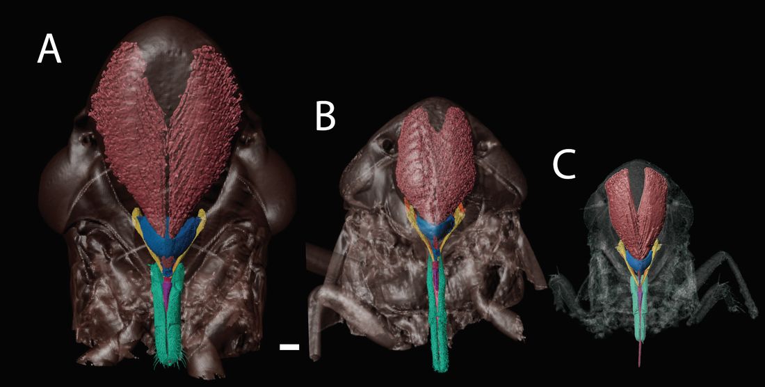
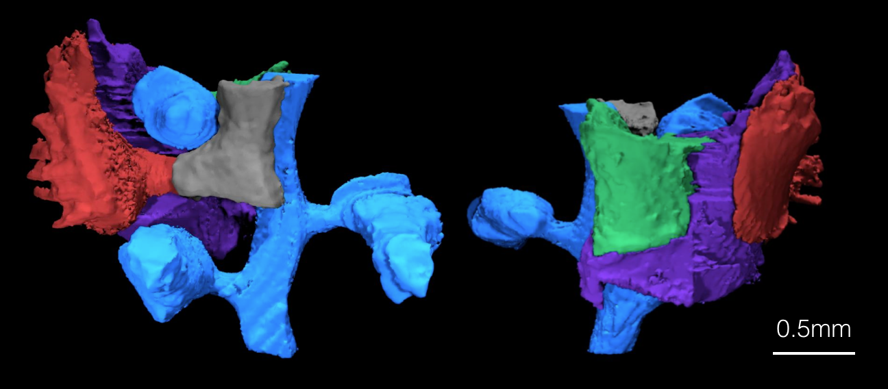
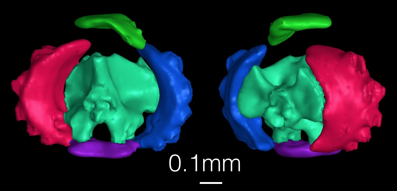
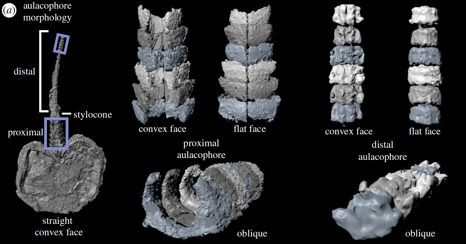
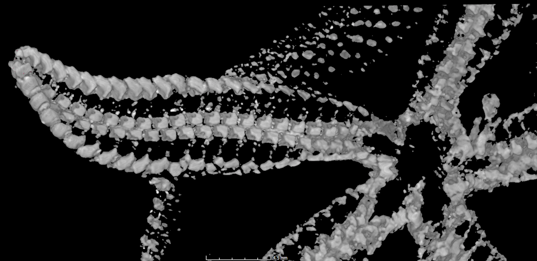
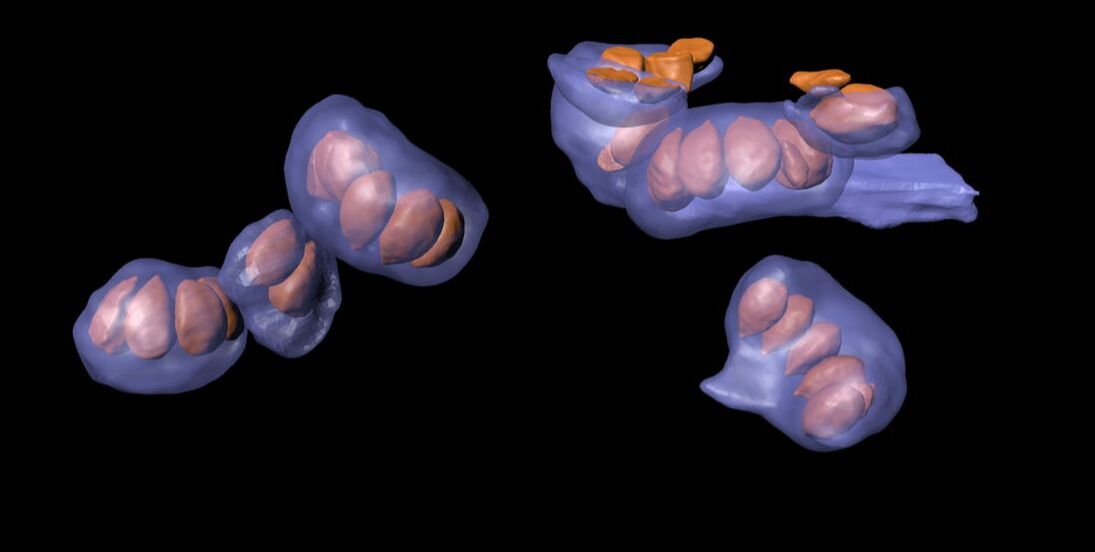
There is a group of insects, the Auchennorynchans, that feed on the xylem sap of plants. These insects are known to transmit bacterial pathogens that cause lethal diseases in important crops in the process of feeding on them. In California, the glassy winged sharpshooter (Homalodisca vitripennis) (A), the meadow spittlebug (Philaenus spumarius) (B) and the blue-green sharpshooter (Graphocephala atropunctata) (C) transmit pathogens to grapes, almonds, citrus fruit, blueberries and more. There is a lot we don't understand about xylem sap-feeding that's necessary to understand how pathogens are transmitted in this process. These 3D reconstructions show the anatomy of the feeding complex, which is helping us to better understand how their anatomy facilitate feeding and pathogen transmission.
Clark et al. 2023. Data from: Anatomy of an agricultural antagonist: feeding complex structure and function of three xylem sap-feeding insects illuminated with synchrotron-based 3D imaging. Dryad. https://doi.org/doi:10.5061/dryad.s4mw6m99s |
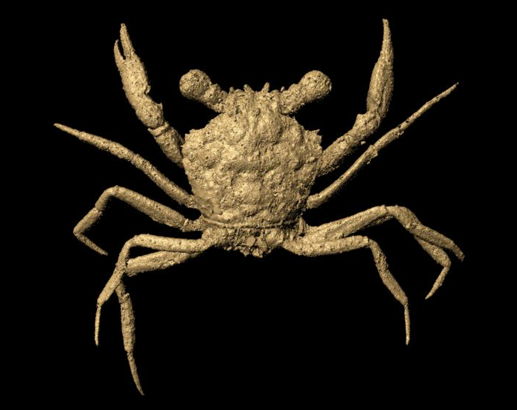
3D Model of Most Complete Fossil Crab Ever Discovered
|
Cretapsara athanata, preserved in 100 million year old amber, is the oldest known modern-looking fossil crab. With gills preserved in tact, this specimen tells the story of how crabs made the transition from the water to terrestrial environments. Luque, J., Xing, L., Briggs, D.E.G., Clark, E.G., Duque, A., Hui, J., Mai, H., McKellar, R.C. 2021. Crabs in amber reveal an early colonization of freshwater during the Cretaceous. Science Advances 7: 1-12. Link to 3D printable model: https://t.co/Cdl23RiSZO?amp=1 |
3D Images and Digital Models of an Ancient Vascular Plant
|
Exceptionally-preserved fossil clubmoss Lycopodicaulis oellgaardii illuminates the evolutionary history of this group of early vascular plants. The vascular structures in this fossil are even preserved in 3D! Data set includes micro-CT scans, Maya models and 3D object files. Herrera, F., Testo, W. L., Field, A. R., Clark, E. G., Herendeen, P. S., Crane, P. R. 2022. Data from: A permineralized Early Cretaceous lycopsid from China and the evolution of crown clubmosses. figshare. 10.6084/m9.figshare.14925369 |
3D Images and Digital Models of Protasterina flexuosa (CMC 25001)
|
3D images and processed 3D data files of fossil brittle star Protasterina flexuosa (CMC 25001) published in Clark et al. 2017 in Biology Letters. Data set includes Micro-CT scans, Maya models, and STL files. Clark, E. G. et al. 2017. Data from: Water vascular system architecture in an Ordovician ophiuroid. Dryad Digital Repository. https://doi.org/10.5061/dryad.30k44 |
3D Images and Digital Models of Ophiothrix angulata and Ophioderma brevispina
|
3D images and processed data from arms of extant brittle stars Ophiurina lymani and Ophioderma brevispina published in Clark et al. 2018 in Journal of Anatomy. Data set includes Micro-CT scans and Maya models.
Clark, E. G. et al. 2018. Data from: Integrating morphology and in vivo skeletal mobility with digital models to infer function in brittle star arms. Figshare. https://figshare.com/projects/Data_from_Integrating_morphology_and_in_vivo_skeletal_mobility_with_digital_models_to_infer_function_in_brittle_star_arms/37709 |
3D Images of Ophiarachna incrassata
|
3D imaging data from extant brittle stars Ophiarachna incrassata published in Clark et al. 2019 in Journal of Experimental Biology. Data set includes Micro-CT scans and video files from behavioral experiments.
Clark, E. G. et al. 2019a. Data from: The function of the ophiuroid nerve ring: How the decentralized nervous system controls locomotion. Dryad Digital Repository. https://doi.org/10.5061/dryad.pv814j1 |
Phase-Contrast Synchrotron Images of Ophioderma brevispina
|
Phase-contrast synchrotron x-ray images from extant brittle stars Ophioderma brevispina published in Clark et al. 2019 in Zoomorphology. Clark, E. G. et al. 2019b. Data from: A farewell to arms: using X-ray synchrotron imaging to investigate autotomy in brittle stars. Dryad Digital Repository. http://doi.org/10.6084/m9.figshare.8049890.v1 |
3D Images and Digital Kinematic Models of Fossil Stylophoran
|
3D imaging data from fossil stylophoran Phyllocystis crassimarginata published in Clark et al. 2020 in Royal Society Open Science. Data set includes Micro-CT scans and range of motion models in Maya.
Clark, E. G. et al. 2020. Data from: Arm waving in stylophoran echinoderms: three-dimensional mobility analysis illuminates cornute locomotion. Dryad Digital Repository. http://doi.org/10.5061/dryad.3sv28v7 |
3D Images and Digital Models of Fossil Ophiuroids from the Devonian Hunsrück Slate
|
3D images of six fossil brittle stars from the Devonian Hunsrück Slate: Encrinaster roemeri, Euzonosoma tischbeinianum, Loriolaster mirabilis, Cheiropteraster giganteus, Furcaster palaeozoicus, and Ophiurina lymani. Data set includes micro-CT scans and 3D Maya models. Clark, E. G. et al. 2020. Data from: Three-dimensional morphology and locomotion of ophiuroids from the Devonian Hunsrück Slate. Figshare. https://doi.org/10.6084/m9.figshare.11886573.v1 |
3D Images and Digital Models of Angiosperm Ancestors (Angiophytes)
|
3D images and 3D reconstructions of fossil angiosperm ancestors (angiophytes) from the Early Cretaceous of Inner Mongolia (about 125.6 million years ago). Data set includes micro-CT scans and 3D Maya models.
Shi, G., Herrera, F., Herendeen, P. S., Clark, E. G., Crane. P. R. 2021. Data from: Mesozoic cupules and the origin of the angiosperm second integument. Dryad Digital Repository: https://doi.org/10.5061/dryad.5x69p8d2r |